Hello ASPS members,
In this short edition of Phytogen we have a research profile from ASPS member Dr Francine Perrine-Walker (The University of Sydney). See below.
Rare gardening and botanical books for sale!
An expert gardener’s 50 yr lifetime collection of gardening books is for sale, the entire collection is worth around $6,000.
It includes rare and specialist horticultural and botanical books. Some of these are out of print and simply no longer available. Others are leather bound etc. There are also stunning coffee table picture books. The seller, Graeme, would prefer not to sell the books individually, but in large ‘chunks’ or as a complete set, as much is possible. Graeme is asking $3,500 for the entire gardening book collection, or for offers for parts of it. The books can be collected from Alphington, VIC 3078.
The final list and prices for the sale of this magnificent gardening book collection can be accessed HERE.
The list is also available as a google doc if the file is too large.
If you are interested in any of these books please contact Liz Boulton, 0407072480 or liz_boulton@hotmail.com
We are hoping these can go to someone who will obtain genuine value from them.
Research Profile: Dr Francine Perrine-Walker, The University of Sydney.
Investigating Plant-Microbe Interactions: A plant scientist’s stumbling guide using fluorescent stains and microscopy
Where does one begin when investigating the infection process of plant pathogens? My project investigated the interaction of the plant pathogenic oomycete, Phytophthora palmivora, with various horticultural crops such as cocoa.


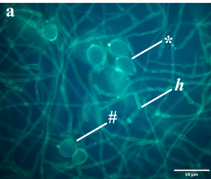
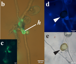
Phytophthora as a plant pathogen resembles true fungi as it forms hyphae (h) and spores as shown in Fig. (a and b).
In its asexual stage, Phytophthora sp. forms chlamydospores (#) which are thick-walled, long-term survival spores and sporangia (*) where short-lived, one-celled, flagellated zoospores are kept safe until fully developed for release in the presence of water.
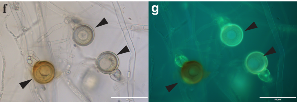
In its sexual stage, the pathogen forms oospores (white and black arrowheads in Figs d-g) resulted from fertilization of the oogonium (female organ) by an antheridium (male organ). Some species are homothallic (self-fertile) while others are heterothallic i.e., requiring cross fertilization.
The marvel of this pathogen? Unlike true fungi, its cell walls contain cellulose instead of chitin! How does one tell them apart when infecting a plant cell?
In the absence of transformed lines with fluorescent tags such as the Green Fluorescence Protein, basic techniques can still shed some light on this pathogen’s cell wall structures. By growing them on agar plates and mating cultures, fluorescent stains such as Calcofluor White (CW) and Aniline Blue (AB) can be used to detect β-1,4-glucans such as cellulose and chitin (Fig.d) and
β-1,3-glucans (Figs. a, b, c and g) respectively. Note when stained with CW, cell wall structures appear blue while with AB, they appear yellow under fluorescence respectively. The latter is the most abundant form of β-glucans in the cell walls of plant
fungal pathogens and, β-1,3-glucans have been shown to act as MAMPs (microbe-associated molecular patterns)
and play a role in plant immune responses.
However, such methods cannot describe the dynamic changes in cell wall content during infection. This requires cutting edge molecular tools other than GFP in combination with high-end microscopy.
 Dr Francine Perrine-Walker (The University of Sydney)
